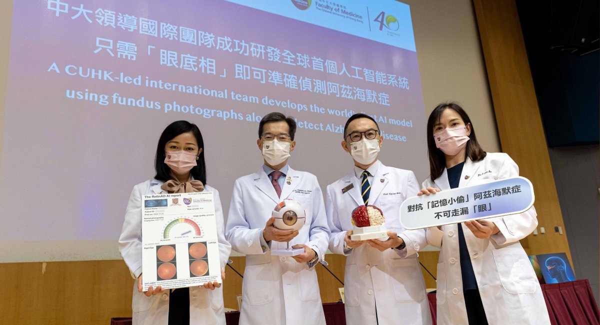
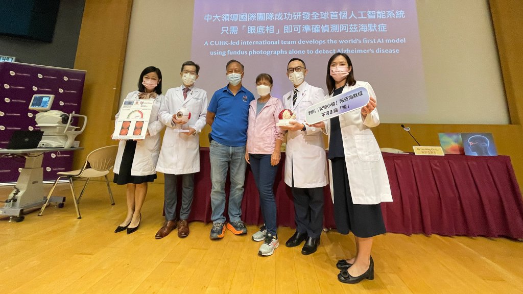
Dr Carol YL CHEUNG, Associate Professor in the Department of Ophthalmology and Visual Sciences at The Chinese University of Hong Kong (CUHK)′s Faculty of Medicine (CU Medicine) has led an international team to successfully develop the world′s first artificial intelligence (AI) model that can detect Alzheimer′s disease solely through fundus photographs or images of the retina. The model is more than 80% accurate after validation.
The retina is an extension of the central nervous system; thus, the eye is therefore a window that can show degenerative changes in the blood vessels and nerves of the brain. Considering fundus photography is widely accessible, non-invasive and cost-effective, this novel AI model incorporated with fundus photography is expected to become an important tool for screening people at high risk of Alzheimer′s disease in the community. Details have been published in The Lancet Digital Health under the international journal The Lancet.
Current methods to detect early Alzheimer′s disease are limited
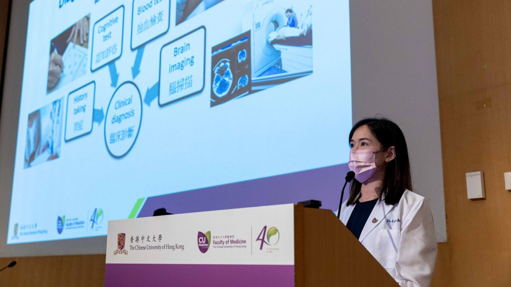
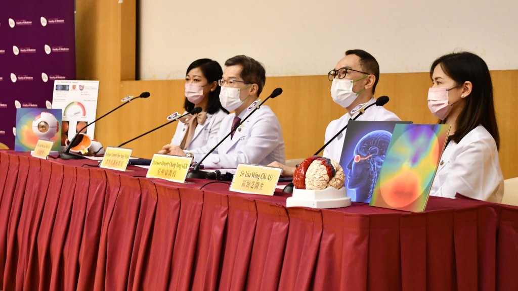
Dr Lisa WC AU, Clinical Professional Consultant of the Division of Neurology in CU Medicine′s Department of Medicine and Therapeutics, said, ‶Memory complaints are common among middle-aged and elderly people, and often considered a sign of Alzheimer′s disease. It is sometimes difficult to make an accurate diagnosis of Alzheimer′s disease based on cognitive tests and structural brain imaging. However, methods to detect Alzheimer′s pathology, such as an amyloid-PET scan or testing of cerebrospinal fluid collected via lumber puncture, are invasive and less accessible.″
To address the current clinical gap, CU Medicine has led a number of medical centres and institutions from Singapore, the United Kingdom and the United States to successfully develop an AI model using state-of-the art technologies which can detect Alzheimer′s disease using fundus photographs alone.
The retina is a window to study disorders of the central nervous system
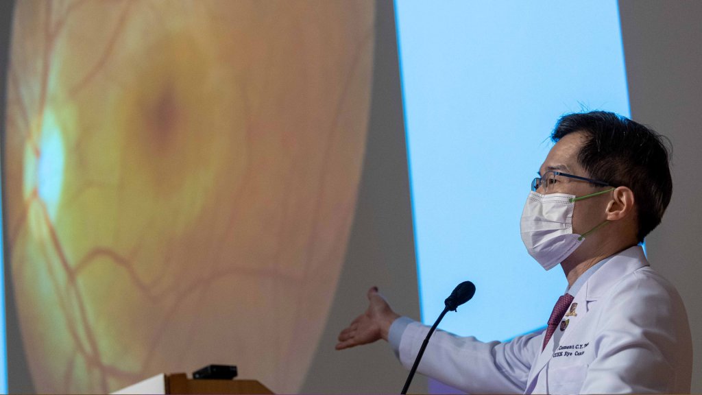
Prof Clement CY THAM, S.H. Ho Professor of Ophthalmology and Visual Sciences and Chairman of CU Medicine′s Department of Ophthalmology and Visual Sciences, explained, ‶The retina is an extension of the brain in terms of embryology, anatomy and physiology. In the entire central nervous system, only the blood vessels and nerves in the retina allow direct visualisation and analysis. Hence, it has long been considered a window to study disorders in the central nervous system. Through non-invasive fundus photography, we can detect a range of changes in the blood vessels and nerves of the retina that are associated with Alzheimer′s disease.″
The team developed and validated their AI model using nearly 13,000 fundus photographs from 648 Alzheimer′s disease patients (included patients from the Prince of Wales Hospital) and 3,240 cognitively normal subjects. Upon validation, the model showed 84% accuracy, 93% sensitivity and 82% specificity in detecting Alzheimer′s disease. In the multi-ethnic, multi-country datasets, the AI model achieved accuracies ranging from 80% to 92%.
Accessibility, non-invasiveness and high cost-effectiveness of the AI model using fundus photography help detection of Alzheimer′s cases both in clinic and the community

Prof Vincent CT MOK, Mok Hing Yiu Professor of Medicine and Director of the Therese Pei Fong Chow Research Centre for Prevention of Dementia at CU Medicine, remarked, ‶In addition to its accessibility and non-invasiveness, the accuracy of the new AI model is comparable to imaging tests such as magnetic resonance imaging (MRI). It shows potential to become not only a diagnostic test in clinics, but also a screening tool for Alzheimer′s disease in community settings. In the future, we hope to validate its efficacy in identifying high-risk cases of the disease hidden in the community, so that various preventive treatments such as anti-amyloid drugs can be initiated early to slow down cognitive decline and brain damage.″
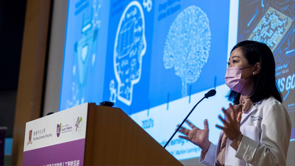
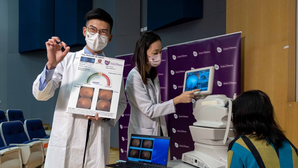
Dr Carol YL CHEUNG, Associate Professor in the Department of Ophthalmology and Visual Sciences at CU Medicine, added, ‶In addition to applying novel AI technologies in the model, we also tested it in different scenarios. Notably, our AI model retained a robust ability to differentiate between subjects with and without Alzheimer′s disease, even in the presence of concomitant eye diseases like macular degeneration and glaucoma which are common in city-dwellers and the older population. This further supports the concept that our AI analysis of fundus photographs is an excellent tool for the detection of the memory-depriving Alzheimer′s disease. To move this research towards clinical application, we are developing an integrated, AI-based platform to combine information from both blood vessels and nerves of the retina captured by fundus photography and optical coherence tomography for detection of Alzheimer′s disease. Our findings should provide more evidence to move AI from code to the real world.″
張艷蕾博士領導團隊成功研發全球首個人工智能系統
只需「眼底相」即可準確偵測阿茲海默症
香港中文大學(中大)醫學院眼科及視覺科學學系副教授張艷蕾博士領導一個國際團隊,成功研發全球首個人工智能系統,只需透過分析「眼底相」(視網膜圖像)便能夠偵測阿茲海默症,準確度逾80%。視網膜是中樞神經系統的延伸,可作為「窗口」反映腦內血管和神經的退化程度。有關系統不但容易應用、非入侵性,且成本效益高,有望日後在社區成為篩查罹患阿茲海默症高危人士的重要工具。相關內容已於國際期刊《刺針》旗下的《The Lancet Digital Health》發表。
目前檢測早期阿茲海默症的方法有限
本港每十名70歲或以上長者便有一位罹患認知障礙症,而患者當中超過一半屬阿茲海默症。他們的腦內過度積聚不正常的物質,包括β-類澱粉狀蛋白及神經纖維纏結,導致腦細胞逐漸死亡,從而引致認知能力退化。
中大醫學院內科及藥物治療學系腦神經科臨床專業顧問區頴芝醫生表示:「中年人及長者出現記憶力衰退非常普遍,亦常被認為是罹患阿茲海默症的徵兆。然而,依據認知測試和腦結構影像掃描來準確診斷阿茲海默症有時候並不容易。現時偵測澱粉狀蛋白的方法,例如正電子腦掃描或通過腰椎穿刺提取腦脊液等,目前仍不普及或具入侵性。」為解決臨床上的限制,中大醫學院率領來自新加坡、英國和美國的多間醫療中心及院校,成功研發這套應用尖端人工智能技術,僅透過「眼底相」已可偵測阿茲海默症的人工智能系統。
視網膜被視為觀察中樞神經系統疾病的平台
中大何善衡眼科及視覺科學講座教授兼中大醫學院眼科及視覺科學學系系主任譚智勇教授解釋:「視網膜一向被視為大腦的延伸,兩者在胚胎學、生物特徵和結構上非常相似。眼底內的血管和視覺神經更是中樞神經系統中唯一可以直接觀察和分析的血管和神經線,能作為研究中樞神經系統疾病的平台。因此,藉着拍攝『眼底相』這種非入侵性的方法,我們可以研究眼底血管或視覺神經有否出現和阿茲海默症相關的病變。」
研究團隊利用近13,000張來自648名阿茲海默症患者(包括威爾斯親王醫院的病人)和3,240名認知功能正常人士的「眼底相」,用於研發和測試新系統。經實驗驗證,新系統偵測阿茲海默症的準確度為84%,敏感度和特異性分別達93% 和82%。另外,將新系統應用於來自不同種族和國家的「眼底相」數據,準確度亦達80%至92%。
檢測方法簡易、無創、安全及低成本 有助從社區和診所揪出阿茲海默症患者
中大莫慶堯醫學教授、中大周佩芳認知障礙預防研究中心主任莫仲棠教授指出:「新系統的準確度可媲美磁力共振等影像檢查,而且容易應用、不具入侵性。它不僅有潛力應用在診所協助診斷阿茲海默症,更可成為在社區進行相關疾病篩查的工具。日後,我們有望以『眼底相』配合人工智能系統,便能找出隱藏在社區的阿茲海默症高危個案,盡早為這些患者進行各種預防治療,例如使用可抑制澱粉狀蛋白積聚的藥物,以減慢他們的認知功能退化及減低對腦部的損害。」
中大醫學院眼科及視覺科學學系副教授張艷蕾博士補充:「我們在新系統中應用了尖端的人工智能技術,並已測試其在不同情況下的效用。即使接受檢查人士患有黃斑病變和青光眼這些都市人和長者較常見的眼科疾病,我們的人工智能系統仍能有效偵測阿茲海默症相關的視網膜特徵,猶如讀取阿茲海默症的生物指紋。因此,如要對抗阿茲海默症這個記憶小偷,切記不可走漏『眼』。我們現正開發一個人工智能綜合系統,可以結合眼底相和光學相干斷層掃描中有關眼底血管和神經線的病變資訊,用作檢測阿茲海默症。有關研究結果將為人工智能系統在阿茲海默症偵測的臨床應用提供可靠數據,將人工智能技術融入我們生活當中。」
 Creative Commons Attribution